A confocal microscope is a type of microscopy that uses a focused laser beam to illuminate a sample and a pinhole aperture to eliminate out-of-focus light, resulting in high-resolution images with improved contrast and clarity.. The name “confocal” refers to the use of a pinhole aperture to achieve optical sectioning of the specimen.
Confocal microscopy was first invented by Marvin Minsky in 1957. At the time, Minsky was a graduate student at Harvard University working on the development of a microscope that could provide high-resolution images of thick biological specimens. He proposed a new type of microscope that would use a pinhole aperture to eliminate out-of-focus light, allowing for sharper and clearer images of thick specimens.
However, it was not until the 1980s that confocal microscopy became widely used, thanks to the development of better lasers, computer-controlled scanning systems, and high-sensitivity detectors. Today, confocal microscopy is an essential tool in many fields of science, including biology, medicine, and materials science. It has enabled researchers to study complex biological systems in unprecedented detail, leading to new insights into the structure and function of living organisms. Figure 1 shows a schematic of the system.
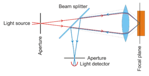
Figure 1.Confocal Microscopy. Image from wikipedia
Key Components of Confocal Microscope
Confocal microscopy is a complex imaging technique that involves several key components. These components work together to produce high-resolution images of thick biological specimens with improved contrast and clarity. Some of the major components used in confocal microscopy include:
- Laser: Confocal microscopes use lasers as the light source for illumination. The laser provides a focused beam of light that can be precisely controlled in terms of its wavelength, intensity, and polarization.
- Scanning system: The laser beam is directed onto the specimen through a scanning system, which typically consists of a pair of mirrors that can rapidly deflect the beam in two dimensions. This allows the beam to be scanned across the specimen in a controlled and precise manner.
- Microscope objective: The laser beam is focused onto the specimen through a microscope objective lens, which collects and focuses the light reflected or emitted by the specimen.
- Pinhole aperture: After passing through the objective lens, the reflected or emitted light is passed through a pinhole aperture. This aperture blocks all light that is out of focus with the objective lens, resulting in optical sectioning of the specimen and improved resolution.
- Detector: The light that passes through the pinhole aperture is detected by a photomultiplier tube or other sensitive detector, which converts the light into an electrical signal that can be processed and displayed as an image.
- Image processing and analysis software: The electrical signal generated by the detector is processed and analyzed using specialized software. This software can be used to enhance contrast, remove noise, and generate 3D images of the specimen.
One of the great advantages of confocal microscopy is the ability to create 3D sample images. Confocal microscopy is particularly useful for imaging thick specimens or samples with complex structures, such as biological tissues or 3D cell cultures.
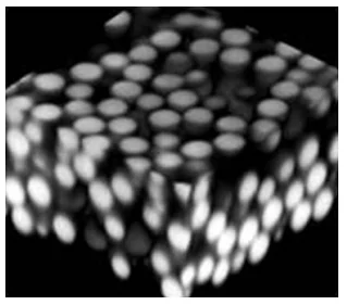
By eliminating out-of-focus light, it is possible to obtain high-resolution images of specific layers or regions of the sample, allowing for detailed analysis of the structure and function of biological systems. Fig. 2 shows a 3D reconstruction from slices of a suspension of 2 mm diameter PMMA particles.
A confocal microscope provides a significant imaging improvement over conventional microscopes. It creates sharper, more detailed 2D images, and allows collection of data in three dimensions.
